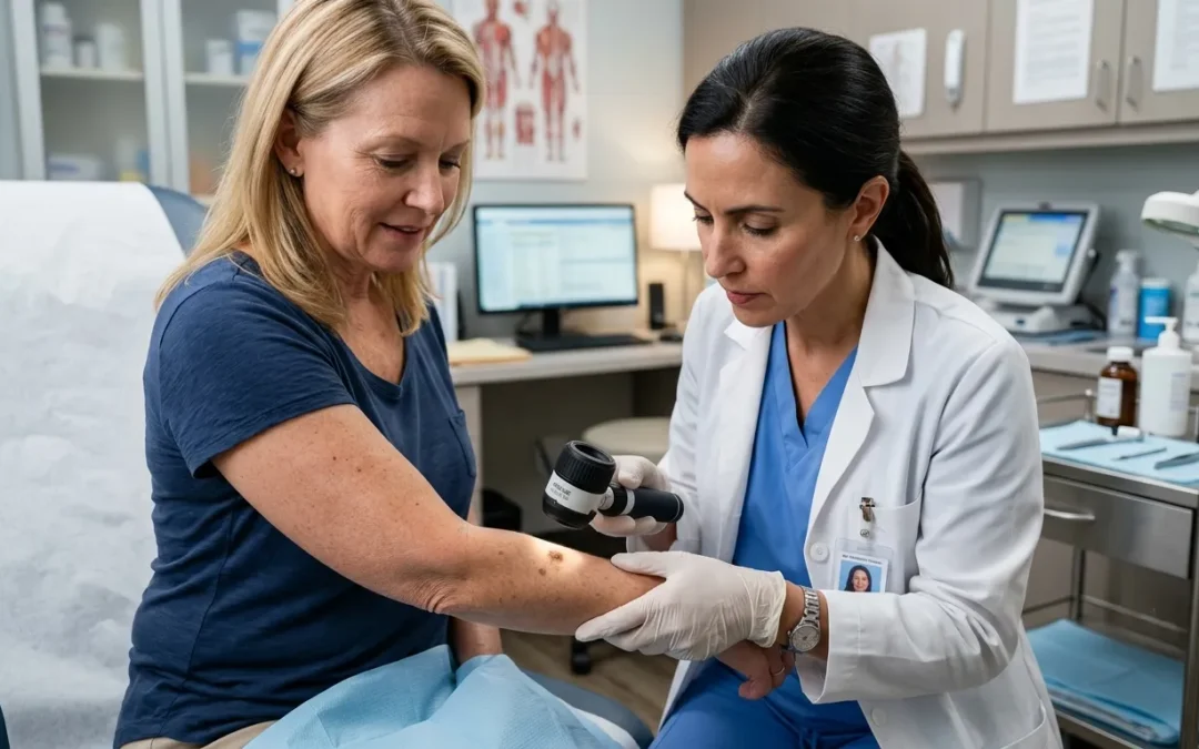
What Does Skin Cancer Look Like? The Ultimate Visual Guide
What Does Skin Cancer Look Like?
Quick Answer
Skin cancer can look like a changing mole, a pearly bump, a scaly patch, a non-healing sore, or a new skin growth. The appearance depends on the type of skin cancer, its stage, and its location on the body. Because there is no single appearance that identifies skin cancer, any persistent or changing skin lesion should be evaluated promptly by a dermatologist.
Table of Contents
Key Takeaways
- Skin cancer does not have one specific appearance.
- A new or changing mole is not the only warning sign.
- Non-healing sores, pearly bumps, and scaly patches can also indicate skin cancer.
- The ABCDE Rule helps identify suspicious pigmented lesions.
- Early recognition improves the likelihood of successful treatment.
One of the most common questions people ask after discovering an unusual spot on their skin is:
"What does skin cancer look like?"
The answer is more complicated than many people expect.
Unlike some medical conditions that have a single recognizable appearance, skin cancer can look very different from one person to another. Some skin cancers resemble harmless pimples, while others appear as persistent sores, rough patches, shiny bumps, or changing moles.
This wide variation explains why skin cancer is sometimes mistaken for:
- Acne
- Eczema
- Psoriasis
- Warts
- Age spots
- Scars
- Minor skin injuries
Although many suspicious skin changes ultimately prove to be harmless, certain lesions require prompt medical evaluation because early-stage skin cancer is often easier to treat than advanced disease.
Learning to recognize the visual warning signs does not mean attempting to diagnose yourself. Instead, it helps you identify changes that deserve professional assessment.
According to the American Academy of Dermatology (AAD) and the American Cancer Society (ACS), becoming familiar with your own skin and recognizing new or evolving lesions plays an important role in early detection.
This guide explains the most common ways skin cancer may appear, how different types of skin cancer can look, and which warning signs should never be ignored.
Does Skin Cancer Always Look the Same?
The answer is No.
One of the biggest misconceptions about skin cancer is that it always appears as a dark or irregular mole. In reality, skin cancer can develop in many different forms depending on:
- The type of skin cancer
- The stage of disease
- The affected skin cells
- Individual skin tone
- The location on the body
Some lesions are:
- Brown
- Black
- Pink
- Red
- White
- Flesh-colored
- Translucent
Others may have little or no pigmentation at all.
For this reason, appearance alone cannot confirm or rule out skin cancer.
What Experts Say?
There is no single "classic" skin cancer appearance. Dermatologists evaluate multiple characteristics—including shape, color, texture, growth pattern, and changes over time—rather than relying on color alone.
Why Skin Cancer Can Be Difficult to Recogniz?
Skin cancer often develops gradually.
During its earliest stages, it may resemble common, non-cancerous skin conditions.
For example, a suspicious lesion may initially appear like:
- A small pimple
- A dry patch
- A tiny cut
- A bug bite
- A wart
- A scar
- A persistent patch of eczema
Because these conditions are common, many people delay seeking medical evaluation until the lesion becomes larger or begins bleeding.
Unfortunately, delaying evaluation can allow certain skin cancers—particularly melanoma—to become more advanced.
Why Visual Changes Matter?
Dermatologists pay close attention to lesions that:
- Continue growing
- Change color
- Develop irregular borders
- Bleed repeatedly
- Fail to heal
- Become crusted
- Persist for several weeks
A lesion that changes over time is often more concerning than one that has remained unchanged for many years.
Common Visual Warning Signs of Skin Cancer
Although skin cancer can look different in every person, several features are commonly associated with suspicious lesions.
These include:
- A new mole appearing in adulthood
- A mole that changes in size or color
- A sore that does not heal
- A shiny or pearly bump
- A rough, scaly patch
- A lesion that repeatedly bleeds
- Crusting or ulceration
- Persistent redness
- A waxy scar-like area
- A firm skin-colored nodule
Not every lesion with these features is cancerous.
However, persistent or changing skin lesions should always be evaluated by a healthcare professional.
Changes That Should Never Be Ignored
Contact a dermatologist if you notice:
- A mole growing rapidly
- Bleeding without injury
- Persistent itching
- Recurrent crusting
- Pain developing within a lesion
- A sore present for more than several weeks
- New pigmentation around an existing mole
Did You Know?
Many early skin cancers are completely painless. Waiting for pain before seeking medical attention may delay diagnosis and treatment.
The ABCDE Rule for Identifying Suspicious Moles
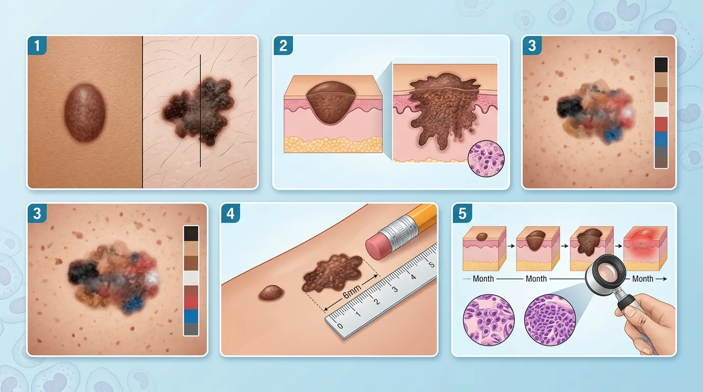
The ABCDE Rule is one of the most widely used tools for recognizing suspicious pigmented lesions that may represent melanoma.
Although it does not diagnose skin cancer, it helps identify changes that warrant professional evaluation.
A — Asymmetry
One half of the mole does not match the other half.
Healthy moles are usually symmetrical.
B — Border
Look for borders that are:
- Uneven
- Blurred
- Notched
- Ragged
Irregular edges may indicate abnormal growth.
C — Color
Healthy moles usually have one consistent color.
Suspicious lesions may contain multiple shades, including:
- Brown
- Black
- Red
- White
- Blue
- Gray
D — Diameter
Many melanomas are larger than 6 millimeters, approximately the size of a pencil eraser.
However, some melanomas are smaller, so size alone should not determine whether a lesion is concerning.
E — Evolving
This is often considered the most important warning sign.
Any mole or skin lesion that changes in:
- Size
- Shape
- Color
- Elevation
- Texture
- Symptoms
should be evaluated promptly.
What Experts Say?
Dermatologists often consider evolution over time to be the strongest predictor of a potentially concerning lesion. Even a small mole deserves attention if it continues changing.
Skin Self-Awareness Saves Lives
Regular skin self-examinations help you recognize changes earlier.
Become familiar with:
- Existing moles
- Freckles
- Birthmarks
- Pigmented spots
- Areas of previous sun damage
Knowing what is normal for your skin makes it easier to identify new or evolving lesions.
Monthly self-examinations are particularly valuable for individuals with:
- Fair skin
- Numerous moles
- Previous skin cancer
- A family history of melanoma
- Extensive lifetime sun exposure
According to Current Research
Clinical evidence consistently shows that early recognition of suspicious skin changes improves treatment outcomes. While self-examinations cannot diagnose skin cancer, they play an important role in identifying lesions that require professional assessment.
What Does Basal Cell Carcinoma Look Like?
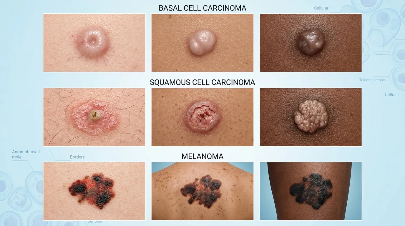
Basal Cell Carcinoma (BCC) is the most common type of skin cancer, accounting for the majority of non-melanoma skin cancers.
It usually develops on areas that receive frequent sun exposure, including:
- Face
- Nose
- Ears
- Neck
- Scalp
- Shoulders
Unlike melanoma, basal cell carcinoma generally grows slowly and rarely spreads to distant organs. However, if left untreated, it can invade nearby skin, cartilage, and bone.
Common Appearance of Basal Cell Carcinoma
Basal cell carcinoma may appear as:
- A shiny or pearly bump
- A flesh-colored or pink nodule
- A sore that repeatedly heals and reopens
- A flat scar-like patch
- A waxy plaque
- A lesion with visible tiny blood vessels (telangiectasia)
Some lesions bleed easily after minor trauma.
Related Guide: Basal Cell Carcinoma: Complete Expert Guide
What Experts Say?
Basal cell carcinoma often appears so harmless that people mistake it for a pimple or minor skin injury. A lesion that repeatedly bleeds or never fully heals should always be evaluated by a dermatologist.
What Does Squamous Cell Carcinoma Look Like?
Squamous Cell Carcinoma (SCC) develops from squamous cells located in the outer layer of the skin.
It commonly appears on areas exposed to sunlight, such as:
- Face
- Lips
- Ears
- Hands
- Forearms
- Scalp
Compared with basal cell carcinoma, SCC has a greater potential to spread if diagnosis and treatment are delayed.
Common Appearance of Squamous Cell Carcinoma
Squamous cell carcinoma may resemble:
- A rough scaly patch
- A thickened plaque
- A crusted growth
- A wart-like lesion
- A persistent ulcer
- A raised red bump
The lesion may:
- Bleed
- Become tender
- Develop a crust
- Fail to heal
Related Guide: Squamous Cell Carcinoma Explained
What Does Melanoma Look Like?
Melanoma develops from pigment-producing cells called melanocytes.
Although it accounts for a smaller percentage of skin cancers, melanoma causes most skin cancer-related deaths because it can spread rapidly to other parts of the body if not detected early.
Common Appearance of Melanoma
Melanoma often appears as:
- A changing mole
- A new dark spot
- An irregular pigmented lesion
- A lesion containing multiple colors
- A mole with blurred borders
Color variations may include:
- Brown
- Black
- Blue
- Gray
- Red
- White
Some melanomas contain very little pigment and may appear pink or flesh-colored.
The "Ugly Duckling" Sign
Dermatologists often use the Ugly Duckling Sign.
If one mole looks noticeably different from all your other moles, it deserves closer attention.
Examples include:
- Darker than surrounding moles
- Larger than other moles
- Different shape
- Different texture
- Rapidly changing appearance
Related Guide: Melanoma Skin Cancer: The Ultimate Expert Guide
Did You Know?
Not every melanoma develops from an existing mole. Many melanomas begin as completely new skin lesions.
What Does Early Skin Cancer Look Like?
One reason skin cancer is frequently diagnosed early is that visible changes often appear before other symptoms develop.
Early skin cancer may resemble:
- A tiny pink bump
- A slightly scaly patch
- A small persistent sore
- A changing mole
- A flat reddish lesion
- A pearly nodule
- A waxy scar-like area
Many early lesions are:
- Small
- Painless
- Slowly growing
Because these changes can seem insignificant, they are sometimes ignored for months.
Related Guide: Early Stage Skin Cancer: Signs, Symptoms & Treatment
What Does Skin Cancer Look Like on the Face?
The face receives more lifetime ultraviolet exposure than almost any other body area.
As a result, it is one of the most common locations for skin cancer.
Frequently affected areas include:
- Forehead
- Nose
- Eyelids
- Cheeks
- Temples
- Lips
- Ears
Facial skin cancer may appear as:
- A shiny bump
- Persistent redness
- Crusted sores
- A scar-like patch
- Thickened skin
- Pigmented lesions
Early diagnosis is especially important because facial treatment aims to preserve both function and appearance.
What Does Skin Cancer Look Like on the Nose?
The nose is particularly vulnerable because it protrudes from the face and receives direct sunlight.
Common warning signs include:
- A sore that never heals
- Repeated bleeding
- Persistent crusting
- Pearly nodules
- Small ulcers
- Red scaly patches
Basal cell carcinoma is especially common on the nose.
Related Guide: Skin Cancer on the Nose: Symptoms, Pictures & Treatment
Can Skin Cancer Look Like a Pimple?
Yes.
Some skin cancers initially resemble:
- Pimples
- Acne bumps
- Ingrown hairs
The difference is that ordinary pimples usually heal within days or weeks, whereas suspicious lesions persist or continue growing.
Seek medical evaluation if a "pimple":
- Never disappears
- Bleeds easily
- Crusts repeatedly
- Continues enlarging
Returns in the same location
Can Skin Cancer Look Like Eczema?
Sometimes.
Certain skin cancers, particularly squamous cell carcinoma, may resemble:
- Dry patches
- Eczema
- Psoriasis
- Chronic dermatitis
However, unlike eczema, these lesions often:
- Continue enlarging
- Bleed
- Develop thick crusts
- Fail to improve with moisturizers or topical corticosteroids
Persistent "eczema" that does not respond to appropriate treatment should be examined by a dermatologist.
Can Skin Cancer Look Like a Scar?
Yes.
Some basal cell carcinomas appear as:
- Flat
- White
- Waxy
- Scar-like plaques
Because these lesions often lack obvious pigmentation, they are sometimes mistaken for old scars.
Can a White Spot Be Skin Cancer?
Most white spots on the skin are not cancerous.
Common non-cancerous causes include:
- Vitiligo
- Fungal infections
- Post-inflammatory pigment loss
- Benign scars
However, certain skin cancers may occasionally present as:
- White plaques
- Pale waxy lesions
- Scar-like growths
A white lesion that:
- Continues enlarging
- Changes appearance
- Bleeds
- Does not heal
should always be evaluated by a healthcare professional.
Common Skin Conditions That May Resemble Skin Cancer
Many harmless conditions resemble skin cancer, including:
- Seborrheic keratosis
- Cherry angioma
- Solar lentigo (age spots)
- Common warts
- Eczema
- Psoriasis
- Acne
- Cysts
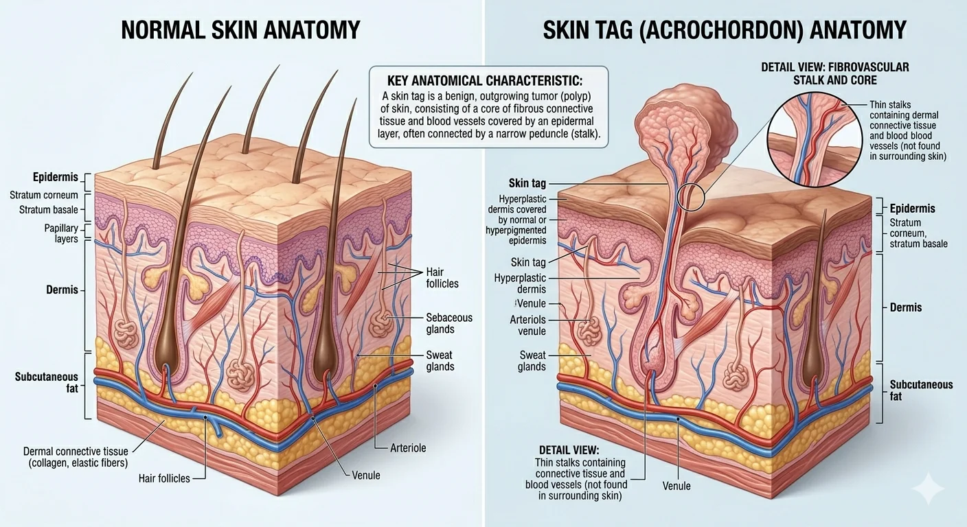
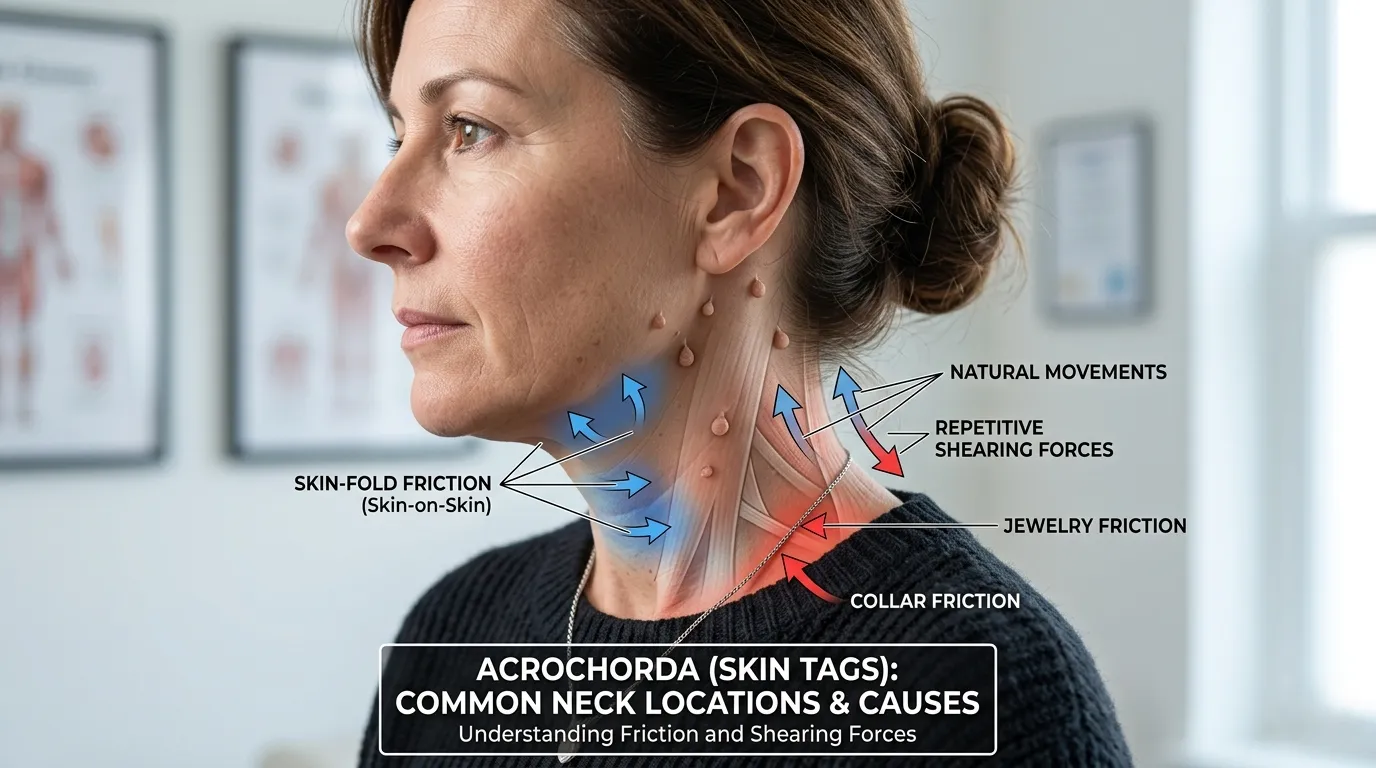
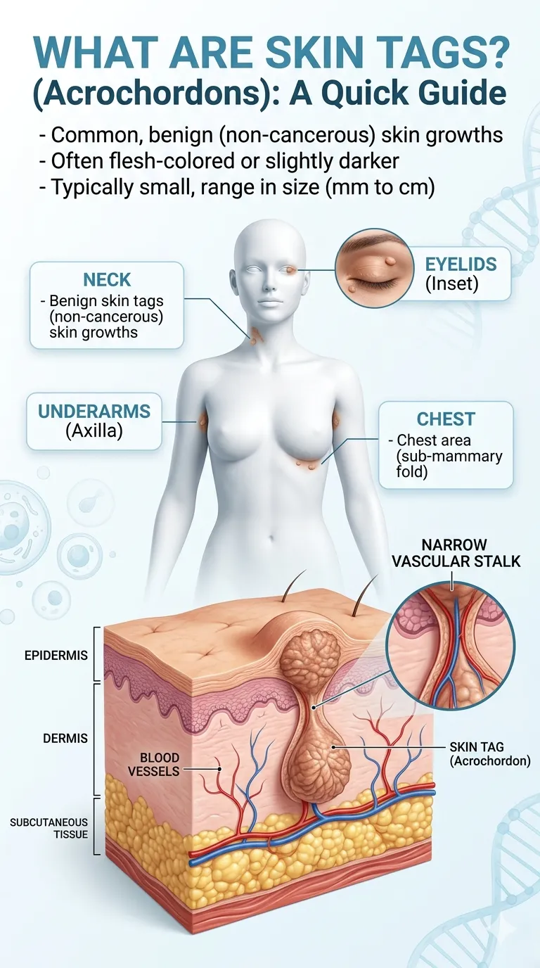
- Skin tags
Because visual appearance alone cannot reliably distinguish these conditions, professional evaluation is essential whenever uncertainty exists.
What Experts Say?
Even experienced dermatologists often use dermoscopy and, when necessary, a skin biopsy to confirm a diagnosis. Self-examination is valuable for identifying suspicious changes, but it cannot replace a professional medical assessment.
When Should You See a Dermatologist?
Not every mole, spot, or skin growth is cancerous, but some changes should never be ignored.
Schedule an appointment with a dermatologist if you notice:
- A new mole appearing in adulthood
- A mole changing in size, shape, or color
- A sore that does not heal within several weeks
- Recurrent bleeding without injury
- Persistent crusting
- A shiny or pearly bump that continues growing
- A rough scaly patch that does not improve
- A lesion that repeatedly heals and returns
- Persistent itching, tenderness, or pain
- Any skin change that concerns you
Early evaluation does not necessarily mean you have skin cancer—it means you are giving yourself the best opportunity for early diagnosis if treatment is needed.
High-Risk Individuals Should Be Especially Vigilant
You may benefit from regular professional skin examinations if you have:
- Fair skin
- Numerous moles
- A personal history of skin cancer
- A family history of melanoma
- Frequent sunburns
- Extensive outdoor sun exposure
- Previous tanning bed use
- A weakened immune system
Individuals with these risk factors should discuss an appropriate skin examination schedule with their dermatologist.
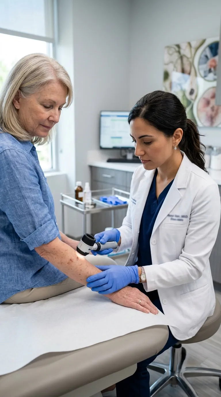
How Dermatologists Diagnose Skin Cancer?
Visual appearance alone cannot confirm whether a lesion is cancerous.
Dermatologists combine clinical expertise with specialized diagnostic techniques to reach an accurate diagnosis.
Clinical Skin Examination
The evaluation begins with a complete skin examination.
Your dermatologist will assess:
- Size
- Shape
- Color
- Border
- Texture
- Location
- Pattern of growth
You may also be asked about:
- Family history
- Previous skin cancers
- Sun exposure
- Tanning bed use
- Recent changes in the lesion
Dermoscopy
A dermoscope is a handheld device that magnifies skin structures invisible to the naked eye.
Dermoscopy helps identify characteristic patterns associated with:
- Melanoma
- Basal cell carcinoma
- Squamous cell carcinoma
- Benign moles
It improves diagnostic accuracy without causing pain.
Skin Biopsy
If a lesion appears suspicious, a skin biopsy is usually recommended.
A small tissue sample is removed and examined under a microscope by a pathologist.
Common biopsy techniques include:
- Shave biopsy
- Punch biopsy
- Excisional biopsy
A biopsy is the gold standard for confirming skin cancer.
Imaging Studies
Most early skin cancers do not require imaging.
However, advanced cancers may require:
- Ultrasound
- CT scan
- MRI
- PET scan
to determine whether cancer has spread beyond the skin.
What Experts Say?
Never attempt to diagnose skin cancer solely from online photographs. Many harmless skin conditions closely resemble skin cancer, while some dangerous cancers initially appear subtle. Only a clinical examination—and, when indicated, a biopsy—can establish a definitive diagnosis.
How to Perform a Monthly Skin Self-Examination?
Monthly self-examinations help you recognize new or changing lesions between professional skin checks.
Choose a well-lit room and use both a full-length mirror and a handheld mirror.
Step 1: Examine Your Face
Carefully inspect:
- Forehead
- Nose
- Eyelids
- Lips
- Ears
- Neck
Step 2: Examine Your Upper Body
Check:
- Chest
- Abdomen
- Back
- Shoulders
- Underarms
Women should also examine the skin beneath the breasts.
Step 3: Examine Your Arms and Hands
Inspect:
- Upper arms
- Forearms
- Palms
- Fingernails
- Between fingers
Step 4: Examine Your Legs and Feet
Check:
- Thighs
- Knees
- Calves
- Soles
- Toes
- Between toes
Step 5: Examine Hard-to-See Areas
Use a handheld mirror or ask a family member to help inspect:
- Back
- Buttocks
- Scalp
- Back of the neck
Photographing suspicious lesions over time can help identify changes, but photographs should never replace professional evaluation.
Myths vs. Facts About Skin Cancer Look Like
| Myth | Fact |
| Skin cancer is always black or dark brown. | Skin cancer may be pink, red, white, flesh-colored, translucent, or pigmented. |
| A painless lesion cannot be skin cancer. | Many early skin cancers cause no pain at all. |
| Only large moles are dangerous. | Small lesions can also represent melanoma or other skin cancers. |
| Skin cancer always develops from an existing mole. | Many skin cancers develop as completely new lesions. |
| Only fair-skinned people develop skin cancer. | People of every skin tone can develop skin cancer. |
| Online pictures are enough to diagnose skin cancer. | Images can educate but cannot replace professional medical evaluation and biopsy. |
Frequently Asked Questions
What does early skin cancer usually look like?
Early skin cancer may appear as a new mole, a changing mole, a non-healing sore, a pearly bump, or a rough scaly patch. The appearance varies depending on the type of skin cancer.
Can skin cancer look like a pimple?
Yes. Some basal cell carcinomas and other skin cancers initially resemble pimples. A "pimple" that persists, bleeds, or repeatedly returns should be evaluated by a dermatologist.
Should every changing mole be checked?
Yes. Any mole or skin lesion that changes in size, shape, color, or texture should be evaluated by a qualified dermatologist.
Can skin cancer look like eczema?
Yes. Certain skin cancers, especially squamous cell carcinoma, may resemble eczema or psoriasis. Persistent patches that do not improve with appropriate treatment require medical evaluation.
What does melanoma usually look like?
Melanoma often appears as a changing mole with asymmetry, irregular borders, multiple colors, or rapid evolution. The ABCDE Rule is a useful tool for identifying suspicious pigmented lesions.
Can skin cancer be white?
Yes. Some basal cell carcinomas appear as white, waxy, or scar-like lesions rather than dark moles.
Expert Summary
Skin cancer has no single recognizable appearance, which is why awareness of changing skin lesions is so important. While some cancers resemble dark moles, others may appear as pearly bumps, persistent sores, scaly patches, scar-like plaques, or even flesh-colored growths.
Rather than trying to diagnose skin cancer yourself, focus on recognizing suspicious changes and seeking timely medical evaluation. Regular self-examinations, routine dermatology visits for individuals at increased risk, and prompt assessment of evolving lesions can significantly improve the chances of early diagnosis and successful treatment.
Conclusion
Recognizing what skin cancer may look like is an important first step in protecting your long-term health.
Although many harmless skin conditions resemble skin cancer, new, changing, or non-healing skin lesions should never be ignored. Learning the ABCDE Rule, performing monthly skin self-examinations, and seeking prompt evaluation from a dermatologist when suspicious changes occur can greatly improve the likelihood of early detection.
Remember that online images and educational articles are valuable for raising awareness, but they cannot replace a professional medical examination. When in doubt, have your skin checked—early action can make a significant difference.
Scientific References
- American Academy of Dermatology (AAD). Skin cancer warning signs, ABCDE rule, and skin self-examination guidance.
- American Cancer Society (ACS). Skin Cancer Prevention, Detection, and Early Recognition.
- National Cancer Institute (NCI). Skin Cancer (Including Melanoma) Patient Information.
- World Health Organization (WHO). Ultraviolet Radiation and Human Health.
- Centers for Disease Control and Prevention (CDC). Skin Cancer Awareness and Prevention.
- National Institutes of Health (NIH). Research on ultraviolet radiation, skin lesions, and carcinogenesis.
- National Library of Medicine (PubMed). Clinical studies on dermoscopy, melanoma detection, and skin cancer diagnosis.
- Journal of the American Academy of Dermatology (JAAD). Research on melanoma recognition, dermoscopy, and skin cancer screening.
- British Association of Dermatologists (BAD). Patient guidance on recognizing suspicious skin lesions.
About the Author
This article was written by Dr. Aamir Raza.
Dr. Aamir Raza is a Phytomedicine Researcher, Herbal Physician, and Evidence-Based Health & Skincare Educator with more than 10 years of university teaching experience. He specializes in phytomedicine, dermatological science, herbal therapeutics, preventive healthcare, and translating peer-reviewed medical research into practical, accurate, and evidence-based health information.
Professional Credentials
🏅 M.Phil. (Phytomedicine) — Gold Medalist
🎓 BEMS (Bachelor of Eastern Medicine & Surgery) — Silver Medalist
👨🏫 10+ Years of University Teaching Experience
🌿 Herbal Physician
🔬 Phytomedicine Researcher
📝 Evidence-Based Health & Skincare Educator
Editorial Review Process
Every article published on Lunaria Organics follows a structured editorial workflow designed to meet Google's EEAT (Experience, Expertise, Authoritativeness, and Trustworthiness) standards.
Our editorial process includes:
- Comprehensive review of current scientific literature.
- Evaluation of peer-reviewed dermatology and oncology research.
- Verification against recommendations from internationally recognized medical organizations.
- Balanced discussion of benefits, limitations, and current scientific evidence.
- Scheduled content reviews to incorporate emerging research.
- Reader-focused educational content that supports informed health decisions.
Medical Disclaimer
The information published on Lunaria Organics is intended for educational and informational purposes only. It should not replace professional medical advice, diagnosis, or treatment. If you notice a suspicious mole, skin lesion, or non-healing sore, seek prompt evaluation from a qualified dermatologist or healthcare professional.